9 Cell voltage
The heart is a pump that contracts in a carefully controlled cycle. Each contraction must be initiated on schedule and in the correct sequence, sixty to a hundred times a minute across a lifetime (Kashou et al. 2022). Beginning the cycle requires a trigger. This is initiated by an electrical signal that originates from a special type of cell in the heart and propagates through surrounding heart cells. Measuring the propagation of this electrical signal provides a detailed view of whether the heart is functioning correctly.
An EKG is the instrument that records the voltage changes between electrodes on the skin. But where does that electrical signal originate? It occurs because a voltage exists across a cell’s membrane. Electrically the cell resembles a small charged battery of a few tens of millivolts, sustained continuously by pumping ions. Most cells hold that voltage steadily. Cardiac muscle cells, however, can also discharge this voltage (depolarize) and trigger the depolarization of neighboring cells. This results in a depolarization wave that propagates through heart tissue.
Cell membrane potential
A voltage, or potential difference, between two points is the energy required to move a unit of electric charge from one point to the other. Its unit is the volt (V), defined as one joule per coulomb. Consider a 1.5-volt AA battery: work has been done to separate positive and negative charge. In general, wherever charge is separated, a voltage exists.
A cell membrane separates charge the same way, on a far smaller scale. The membrane is a lipid bilayer, a film about five nanometers thick, whose oily interior is impermeable to charged ions. In electrical terms it is a thin insulator separating two good conductors: the saline fluid inside the cell (the cytoplasm) and the saline fluid outside. By convention the membrane potential \(V_m\) is defined as the interior voltage minus the exterior,
\[V_m = V_\text{in} - V_\text{out},\]
Defined this way, a resting cell registers a negative value — the interior lies below the exterior by about \(-70\) millivolts (Rye et al. 2016; Chrysafides et al. 2023). Seventy thousandths of a volt is small in absolute terms, but it occurs across only five nanometers of membrane, so the electric field within the film is actually large — strong enough to open the gates of protein channels, which allow charge to flow.
Ion gradients and the pump that maintains them
The charge carriers in a cell are not electrons but ions. Ions are atoms that carry a net charge when dissolved in water. Potassium ions (\(\text{K}^+\)) and sodium ions (\(\text{Na}^+\)) are often the most important for generating the membrane potential. The essential feature is that the cell maintains (\(\text{Na}^+\)) and (\(\text{K}^+\)) at different concentrations inside and outside. The exact levels vary by cell type, but potassium is typically concentrated inside the cell and dilute outside; sodium shows the opposite distribution, abundant outside and low inside (Figure 9.1). Representative values for a mammalian cell are (Rye et al. 2016):
| Ion | Outside (mM) | Inside (mM) |
|---|---|---|
| \(\text{Na}^+\) (sodium) | 145 | 12 |
| \(\text{K}^+\) (potassium) | 4 | 155 |
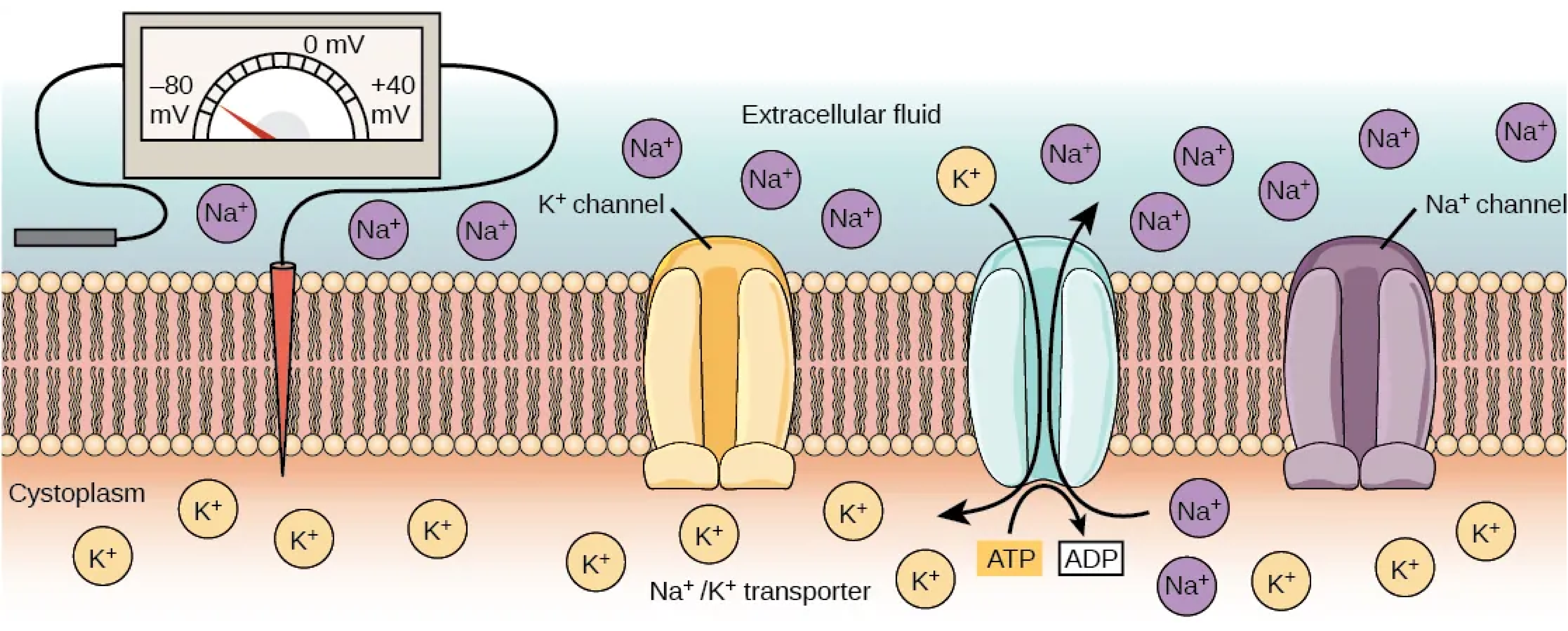
These gradients are sustained by membrane proteins, for example the sodium–potassium pump (\(\text{Na}^+/\text{K}^+\) ATPase). In each cycle, this pump transports three \(\text{Na}^+\) ions from inside to outside and two \(\text{K}^+\) ions from outside to inside, moving every ion against its concentration gradient. This uphill transport requires energy, which the pump obtains by hydrolyzing one molecule of ATP, the cell’s energy currency, per cycle (Rye et al. 2016; Chrysafides et al. 2023). This pump operates continuously, and in active tissue it can consume a large fraction of the cell’s total energy budget. The pump therefore acts as the battery charger: it performs the thermodynamic work of separating sodium from potassium and stores that work as the two concentration gradients.
The resting membrane potential
The membrane is not a perfect seal. It contains ion channels — protein pores, each selective for particular ions. This property is selective permeability. At rest a cell has far more open potassium leak channels than sodium channels, so the resting membrane is much more permeable to \(\text{K}^+\) than to \(\text{Na}^+\) (Rye et al. 2016).
These two conditions together generate the voltage. Potassium diffuses outward down its steep concentration gradient, just as any solute spreads from a region of high concentration to one of low concentration. Each \(\text{K}^+\) that exits carries a positive charge out of the cell, while the large intracellular anions it leaves behind (proteins and phosphates), are too large to pass through the channels and cannot follow. The interior therefore becomes progressively more negative, and the outside more positive, as potassium leaves.
This accumulating negativity opposes the outflow that produces it. As the interior grows more negative, it attracts the positive \(\text{K}^+\) ions more strongly back inward, since opposite charges attract. Potassium is thus subject to two opposing forces: a chemical force directed outward (the concentration gradient) and an electrical force directed inward (the negative interior it is itself creating). Initially the chemical force dominates and potassium continues to leave, but each departing ion strengthens the opposing electrical force. The outflow slows progressively until the two forces balance exactly and the net potassium flux vanishes. The membrane then rests at the voltage at which potassium is in equilibrium (equilibrium potential), which is quoted at about \(-90\) mV in physiology texts (Chrysafides et al. 2023).
The cell’s actual resting potential is slightly less negative, around \(-70\) mV, because the membrane is not permeable to potassium alone. A small sodium current enters through the few open sodium channels, and because sodium is driven inward (by both its concentration gradient and the electrical pull of the negative interior) each entering \(\text{Na}^+\) makes the interior slightly less negative. The resting potential settles at a weighted compromise between potassium’s equilibrium value near \(-90\) mV and sodium’s positive one, landing at about \(-70\) mV and remaining close to potassium’s value because potassium’s permeability dominates (Chrysafides et al. 2023). Combining several ions, each with its own permeability, is the role of the Goldman–Hodgkin–Katz equation, which is not required here but is the exact multi-ion generalization of this single-ion account (Chrysafides et al. 2023).
A cell at rest maintains a voltage across its membrane, interior negative, of about
\[V_m \approx -70\ \text{mV}.\]
It arises because (i) the \(\text{Na}^+/\text{K}^+\) pump maintains a steep \(\text{K}^+\) gradient (high inside) at the cost of ATP, and (ii) the resting membrane is predominantly permeable to \(\text{K}^+\), so potassium diffuses outward until the negative interior it creates opposes the outflow strongly enough to halt it. The resting potential therefore lies near potassium’s equilibrium voltage.
The equilibrium potential: the Nernst equation
The voltage at which an ion’s electrical force exactly cancels its concentration force, so that no net current of that ion flows, is its equilibrium potential (or Nernst potential), \(E_\text{ion}\). It depends only on the ratio of the ion’s concentrations across the membrane, through the Nernst equation:
\[E_\text{ion} = \frac{RT}{zF}\,\ln\!\frac{[\text{ion}]_\text{out}}{[\text{ion}]_\text{in}}.\]
Each symbol has a physical meaning:
- \(R\) — the gas constant
- \(T\) — the absolute temperature, so that \(RT\) is a thermal energy quantifying the ions’ thermal motion and hence the strength of diffusion
- \(F\) — Faraday’s constant, the charge of one mole of elementary charges
- \(z\) — the ion’s charge number (\(z=+1\) for \(\text{Na}^+\) or \(\text{K}^+\); \(z=+2\) for \(\text{Ca}^{2+}\)), so that \(zF\) converts a quantity per mole of ions into one per mole of charge
- \([\text{ion}]_\text{out}\), \([\text{ion}]_\text{in}\) — the ion’s concentration outside and inside the cell
The logarithm of the concentration ratio represents the diffusive contribution. The ratio \([\text{ion}]_\text{out}/[\text{ion}]_\text{in}\) determines that the ion concentrated inside (ratio below 1) has a negative equilibrium voltage, an ion concentrated outside (ratio above 1) a positive one, and an ion distributed evenly (ratio 1, \(\ln 1 = 0\)) requires no voltage to hold it in equilibrium.
At the human body temperature of 37 °C, the combination \(RT/F\) evaluates to about 26.7 mV, and converting from the natural logarithm to the base-10 logarithm raises the prefactor to about 61.5 mV (Chrysafides et al. 2023):
\[E_\text{ion} \approx \frac{61.5\ \text{mV}}{z}\,\log_{10}\!\frac{[\text{ion}]_\text{out}}{[\text{ion}]_\text{in}}.\]
In this form the equation yields a useful rule: for a singly charged ion, each tenfold difference in concentration across the membrane corresponds to about 60 mV. A 10-to-1 ratio gives \(\pm 61.5\) mV (the “\(61.5\) mV per decade” the exercises use), a 100-to-1 ratio gives \(\pm 123\) mV, and so forth. This relation permits a rapid estimate of any equilibrium voltage.
The equation can now be applied to the two ions that establish the resting potential, using the concentrations from Table 9.1. The computation requires only a few lines of code.
import numpy as np
import matplotlib.pyplot as plt
plt.style.use("../../theme/bioe10.mplstyle")

def nernst_mV(c_out, c_in, z=1, T=310.0):
    """Equilibrium (Nernst) potential in mV for a monovalent ion at body temperature."""
    RT_F = 8.314 * T / 96485.0 * 1e3        # RT/F in mV -> 26.7 mV at 310 K
    return (RT_F / z) * np.log(c_out / c_in)

E_K  = nernst_mV(4, 155)                    # potassium: high inside
E_Na = nernst_mV(145, 12)                   # sodium: high outside
print(f"per-decade slope = {8.314*310/96485*1e3*np.log(10):.1f} mV")
print(f"E_K  = {E_K:.0f} mV")
print(f"E_Na = {E_Na:+.0f} mV")

# the Nernst line: equilibrium voltage vs the out/in concentration ratio
ratio = np.logspace(-2.2, 2.2, 400)
fig, ax = plt.subplots(figsize=(7.6, 5.0))
ax.plot(ratio, 61.5 * np.log10(ratio), color="#333333",
        label=r"$E_{ion} = 61.5\log_{10}\frac{[ion]_{out}}{[ion]_{in}}$ mV")
ax.axhline(0, color="#bbbbbb", lw=0.8, zorder=1)
ax.axvline(1, color="#bbbbbb", lw=0.8, ls=":", zorder=1)
ax.axhline(-70, color="#D55E00", ls="--")   # resting potential
ax.scatter([4/155], [E_K], color="#CC79A7", zorder=5)
ax.annotate(f"$E_K$ = {E_K:.0f} mV\n(K$^+$ 4/155)", (4/155, E_K),
            textcoords="offset points", xytext=(14, -30), color="#CC79A7")
ax.scatter([145/12], [E_Na], color="#0072B2", zorder=5)
ax.annotate(f"$E_{{Na}}$ = {E_Na:+.0f} mV\n(Na$^+$ 145/12)", (145/12, E_Na),
            textcoords="offset points", xytext=(-90, 14), color="#0072B2")
ax.text(0.006, -55, "resting potential\n-70 mV", color="#D55E00", va="bottom")
ax.set_xscale("log")
ax.set_xlabel(r"concentration ratio  $[ion]_{out}/[ion]_{in}$")
ax.set_ylabel(r"equilibrium potential  $E_{ion}$  (mV)")
ax.legend(loc="lower right")
plt.show()
Running it prints
per-decade slope = 61.5 mV
E_K  = -98 mV
E_Na = +67 mV
Potassium, concentrated inside at nearly 40 times its outside value, therefore has an equilibrium potential of about \(-98\) mV, while sodium, concentrated outside, lies at about \(+67\) mV. Physiology texts commonly quote \(E_K \approx -90\) mV from slightly different measured concentrations; the exact number depends on the assumed gradient but is always strongly negative (Chrysafides et al. 2023).
Pacemaker cells
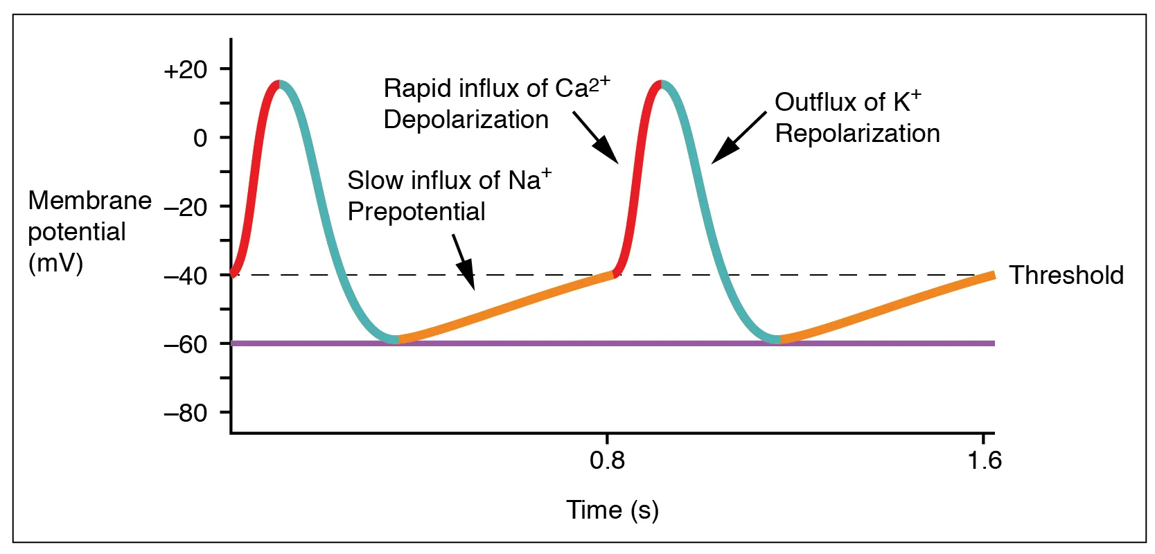
Cardiac cells are excitable: they convert a change in membrane voltage into a large, electrical event, the action potential, driven by a class of ion channels called voltage-gated channel. Once the voltage crosses a threshold, these voltage-gated channels open, ions rush in, and the membrane potential changes rapdily.
A pacemaker cell reaches the triggering potential spontaneously, over and over; a working (contractile) muscle cell waits at rest receiving a push from it’s neighbors.
Pacemaker cells are a small population of specialized cardiac cells that are self-starting. After each action potential their membrane does not settle at a steady resting voltage; instead it immediately begins to depolarize spontaneously, drifting upward until it reaches threshold, at which point it fires another action potential without any external stimulus, and the cycle repeats (Kashou et al. 2022). This slow spontaneous depolarization is the pacemaker potential , shown in Figure 9.2. These cells generate their own rhythm without external timing input.
The heart’s dominant group of these self-firing cells is located in the sinoatrial (SA) node, in the wall of the right atrium. These cells drift to the threshold and fire between 60 and 100 times a minute, which sets the rate of the entire heart. Hence the SA node is termed the heart’s natural pacemaker (Kashou et al. 2022).
Muscle cell action potential
The pacemaker’s signal drives an ordinary working muscle cell to open their voltage-gated channels. Suppose a resting patch of membrane is disturbed so that its voltage rises from \(-70\) mV toward zero — that is, becomes less negative. Once it crosses a threshold of about \(-55\) mV, the voltage-gated sodium channels open. Sodium, which is driven inward throughout (it is concentrated outside and attracted by the negative interior), then flows rapidly into the cell. Each entering \(\text{Na}^+\) carries positive charge inward, raising the voltage further and opening still more sodium channels. This self-amplifying process drives the membrane voltage from \(-70\) mV toward sodium’s equilibrium potential, overshooting to about \(+40\) mV within a fraction of a millisecond. This is called depolarization: the interior briefly becomes positive.
Because the channel opening is self-reinforcing, the spike is all-or-nothing: once threshold is crossed the cell fires completely, independent of the size of the initiating stimulus (Rye et al. 2016). A larger stimulus does not produce a larger spike; it only reaches threshold sooner. The event has a fixed size and shape, which is what makes it a reliable signal.
The sodium channels, having opened, rapidly inactivate — they close and remain shut. But in a cardiac muscle cell the voltage does not then fall straight back to rest. Instead, voltage-gated L-type calcium channels open at the peak of the overshoot and admit a steady inward trickle of \(\text{Ca}^{2+}\) (Gopalan 2021a); because only a few potassium channels are open at the same time, the inward calcium current and the trickle of outward potassium current roughly balance, and the voltage barely moves. This is the plateau: it holds the membrane near its depolarized peak for approximately 175 ms, compared with the mere 3–5 ms the initial depolarization took (Betts et al. 2022). The plateau is what holds the heart muscle contracted long enough to eject blood on each beat, and it stretches the whole electrical event out to a total of roughly 250–300 ms (Betts et al. 2022). These stages are numbered phase 0 (the sodium upstroke), phase 1 (a brief initial notch as sodium channels close), phase 2 (the calcium plateau), phase 3 (repolarization), and phase 4 (rest) (Gopalan 2021a).
Repolarization begins once the calcium channels close: with the inward current gone, potassium efflux dominates unopposed and carries the voltage back down from the plateau to rest, a process that takes a further 75 ms or so (Betts et al. 2022). Because the sodium channels inactivated at the moment of the overshoot and stay shut through the plateau, the cell has an absolute refractory period of about 200 ms during which no stimulus, however strong, can trigger a second spike, followed by a shorter relative refractory period of roughly 50 ms in which only an unusually strong stimulus can (Betts et al. 2022). This refractory period, spanning nearly the entire plateau, caps how soon the cell can fire its next beat. The complete depolarization, plateau, and repolarization is the cardiac action potential, which lasts 250–300 ms from threshold to rest (Betts et al. 2022).
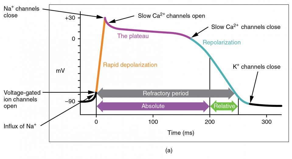
An excitable cardiac cell driven past its threshold (≈ \(-55\) mV) fires an all-or-nothing pulse governed by voltage-gated channels:
- Depolarization, phase 0 (3–5 ms) — voltage-gated \(\text{Na}^+\) channels open, \(\text{Na}^+\) enters, and the voltage rises from \(-70\) mV to about \(+40\) mV (interior briefly positive).
- Plateau, phase 2 (≈175 ms) — voltage-gated L-type \(\text{Ca}^{2+}\) channels open; inward \(\text{Ca}^{2+}\) roughly balances outward \(\text{K}^+\), holding the voltage near its peak (Gopalan 2021a).
- Repolarization, phase 3 (≈75 ms) — \(\text{Ca}^{2+}\) channels close, \(\text{K}^+\) efflux dominates unopposed, and the voltage returns to rest (phase 4), followed by an absolute (≈200 ms) then relative (≈50 ms) refractory period (Betts et al. 2022).
The whole event lasts 250–300 ms.
Propagation: a traveling wave of voltage
A single cell’s action potential (whether self-triggered by a pacemaker or stimulus-triggered in a working muscle cell) is a local event. It propagates from one cell to another because cardiac muscle cells are physically connected to their neighbors by gap junctions. These protein channels link the interior of one cell directly to that of the next. Ions passing through a gap junction carry the depolarization across the cell boundary, so the wave does not stop at a cell’s edge but propagates cell-to-cell through the entire muscle as though it were a single connected wire. Each cell fires as the wave reaches it, and its contraction follows its own action potential shortly after.
The conduction speed sets the timing of the beat. In ordinary heart muscle it is roughly half a meter per second, and along the heart’s specialized fast-conducting fibers (the His–Purkinje system) it reaches a few meters per second. Over the few centimeters of a heart these are modest speeds, which is why the electrical events of one beat are spread over tenths of a second. This islong enough for the EKG to resolve them as distinct deflections. A wave crossing the roughly 8 cm span of the atria at about 1 m/s takes \(0.08\ \text{m} \div 1\ \text{m/s} = 0.08\ \text{s}\), and the atrial deflection on an EKG (the P wave) indeed lasts about that long.
When the SA node fires, the wave of depolarization spreads across the heart, and the next section records the entire sequence at the skin: the wave spreading through the atria (the P wave), a deliberate delay at the junction to the ventricles, the wave passing through the ventricles (the QRS complex), and the ventricles repolarizing afterward (the T wave).This sequence describes a a normal sinus rhythm, which is the signature of a healthy heart.