14 Artificial Skin
A diabetic patient with a foot ulcer may spend months caring for a wound that refuses to close. In a randomized trial of conventional wound care, only 18.3% (21 of 115) of patients with ulcers open longer than six weeks had a fully healed ulcer after 12 weeks (Marston et al. 2003). The engineering solution is to grow a living dermis outside the body, then place it in the wound.
What is skin?
Skin consists of two principal layers. The epidermis is the thin, stratified outer layer composed mainly of keratinocytes; it forms the waterproof barrier but contains no blood vessels. Beneath it lies the dermis, a thicker connective-tissue layer rich in collagen and elastin fibers secreted by fibroblasts, the resident cells of the dermis. The dermis provides mechanical strength, houses sensory receptors and appendages such as hair follicles and sweat glands, and is permeated by a capillary network that supplies oxygen and nutrients to the tissue (Betts et al. 2022).
When skin is damaged, the healing process depends on fibroblasts migrating into the wound bed, proliferating, and rebuilding the collagen matrix. In a chronic diabetic ulcer this process stalls and the matrix signals that normally direct healing are disrupted. Providing a pre-formed dermal matrix seeded with active fibroblasts can improve the healing process.
The tissue-engineering approach
Developed in the 1990s and studied in rigorous clinical trials, DermaGraft is a bioengineered dermal tissue constructed by seeding human neonatal fibroblasts onto a biodegradable polyglactin scaffold and culturing the construct until the fibroblasts deposit a collagen matrix (Marston 2004). The resulting product closely mimics the dermis — the connective-tissue layer beneath the skin surface — and, when applied to a chronic wound, stimulates healing.
Tissue engineering combines living cells with a structural scaffold and a set of biochemical cues that help cells grow. The scaffold serves two purposes: it gives cells a three-dimensional framework to attach to and migrate through, and it eventually degrades as the cells deposit their own matrix. A dermagraft uses a polyglactin mesh (a biodegradable polyester, related to the absorbable sutures used in surgery) as the scaffold. Human neonatal dermal fibroblasts — obtained from neonatal foreskin tissue — are seeded onto this mesh and cultured in a bioreactor for approximately two weeks, during which they proliferate, spread through the mesh, and secrete collagen, fibronectin, and growth factors that together constitute the neodermis — the new dermal tissue the graft builds in place (Marston 2004).
Keeping the dermagraft healthy
We will use the dermagraft as an interesting example in order to start thinking about mass transport. In this case, we are particularly interested in how oxygen diffuses through the scaffold and is consumed by the fibroblasts. However, the approaches that we learn, and the equations we develop, can be applied more broadly to model the movement of solutes.
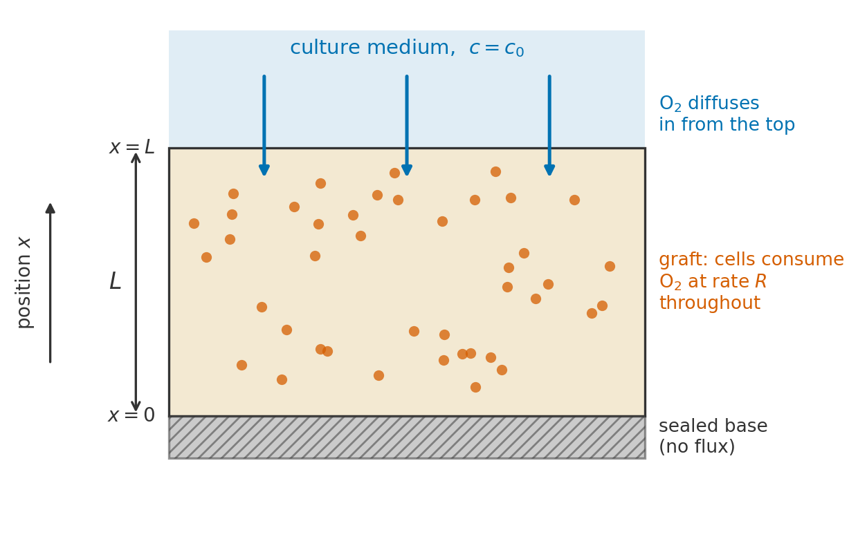
The model reduces the graft to a slab of thickness \(L\) (Figure 14.1). Its top face is in contact with culture medium that holds the oxygen concentration at \(c_0\). Its bottom face rests on a base that oxygen cannot cross. Oxygen enters through the top, diffuses downward, and is consumed by the cells along the way, so the cells at the base are the farthest from the supply. The question of the module is how thick the slab can be before those cells run out of oxygen.
scripts/gen_dermagraft_schematic.py.