37 Introduction
A home COVID-19 antigen test or pregnancy test detects one kind of molecule in a drop of sample. The user adds the sample to a paper strip, waits 5 to 30 minutes, and reads one or two colored lines (Koczula and Gallotta 2016). The strip needs no instrument for a visual read (Andryukov 2020; Koczula and Gallotta 2016).
A biological sample holds thousands of kinds of molecule, and the test must respond to one of them and ignore the rest. The device is the lateral flow assay, or lateral flow test, used in diagnostics for about six decades and suited to point-of-care testing: at the patient’s side, outside a laboratory (Andryukov 2020). How does a lateral flow test turn a few target molecules into a visible line, how long does that take, how little target can it detect, and how often is its result wrong?
Learning objectives
After this module, a student can:
- Fit a calibration line to standards, check the fit with its residuals, and convert a signal into a concentration.
- Find the limit of detection (LoD) from blank readings, compute clinical sensitivity and specificity from a \(2\times2\) table, and compute the positive predictive value at a given prevalence.
- Explain how an antibody recognizes one target, and why a sandwich test needs two antibodies that bind two different parts of the antigen.
- Use the binding rate constants and the dissociation constant \(K_D\) to predict the fraction of antibody bound and the time to a visible line.
- Use viscosity and a force balance to derive the velocity profile in a tube, and compute the flow rate (Poiseuille’s law) and the wall shear stress.
- Compute the capillary pressure in a pore from the surface tension and the contact angle, and the time the sample takes to wick to the test line (the Washburn equation).
- Estimate the time to read a test as the wicking time plus the time to a visible line.
Anatomy of the strip
The strip is a row of overlapping porous pads on a plastic backing (Figure 37.1). Everything it needs is stored dry.
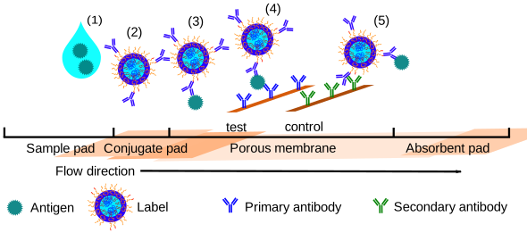

- The drop of sample lands on the sample pad, which meters it onto the strip.
- The conjugate pad holds a dried detection antibody, a protein that binds one specific target molecule (the antigen). Each copy carries a gold nanoparticle, the label, which looks red. The sample redissolves the labeled antibody.
- Antigen in the sample binds the labeled antibody, so the antigen now carries a red label. The labeled antigen then flows onto the nitrocellulose membrane, the porous membrane in the figure.
- The test line, a stripe of capture antibody printed on the membrane, binds the antigen at a second site. A labeled antigen caught there forms a sandwich: capture antibody, antigen, detection antibody. Enough sandwiches make a visible red line. With no antigen in the sample, the line stays blank.
- The control line captures the detection antibody itself, whether or not antigen was present. It must appear in every valid run. If it is missing, the liquid did not flow properly or the dried reagents were spent, and the test line cannot be trusted.
The absorbent pad at the far end soaks up the fluid and keeps it flowing. No pump moves the sample. The dry membrane pulls liquid in by capillary action (wicking), the same effect that draws water into a paper towel.
Before you start
- Rate laws and first-order differential equations: calculus appendix.
- Running code and plotting: Python appendix.
- Mean, spread, and distributions: numerical data.
Roadmap
- Calibration curves: converting a signal to a concentration.
- Diagnostic performance: the limit of detection, and clinical sensitivity and specificity.
- Antibodies: the molecule that recognizes the target.
- Antibody binding: how fast the sensor binds and how long it holds, and from that the time to a visible line.
A sample is detected only if its concentration exceeds both the limit of detection, set by the noise of blank readings (diagnostic performance), and the smallest concentration that gives a visible line, set by the smallest sandwich concentration the eye can see (antibody binding).
The strip moves the sample by capillary flow, and flow in a tube is the model used for each of its pores.
- Flow: viscosity, and the velocity profile of liquid that pressure drives through a tube.
- Poiseuille’s law: the flow rate through a tube, which scales with the fourth power of its radius.
- Capillary flow: what pulls the sample along the strip with no pump, and the total time to read the test.