40  Antibodies

A nasal swab sample holds thousands of kinds of molecule besides the viral protein that a COVID-19 antigen test is built to detect. The test line must turn red when that protein is present and stay blank otherwise. Gold collects at the line when a target molecule links the gold-labeled detection antibody to the capture antibody, so the strip responds to any molecule that both antibodies bind.

An antibody that also bound a protein present in every nasal sample would lack analytical specificity (diagnostic performance). Every test would read positive, so \(TN = 0\) and the clinical specificity, \(TN/(TN+FP)\), would be zero. A lateral flow test therefore needs an antibody that binds the target with negligible binding to other molecules in the sample, has a measured binding strength, and is made to within a stated tolerance from lot to lot (Koczula and Gallotta 2016).

The Y-shaped molecule

An antibody, or immunoglobulin, is a Y-shaped protein. A protein is a chain of amino acids, small molecules linked end to end, folded into a definite shape. An antibody is built from four such chains: two identical heavy chains and two identical light chains. Disulfide bonds, which link sulfur atoms on two amino acids, hold the chains together (Clark et al. 2018). Each chain folds into compact units called domains, a variable domain at its tip and one or more constant domains behind it (Janeway et al. 2001b). IgG, one class of antibody, has this structure (Figure 40.1).
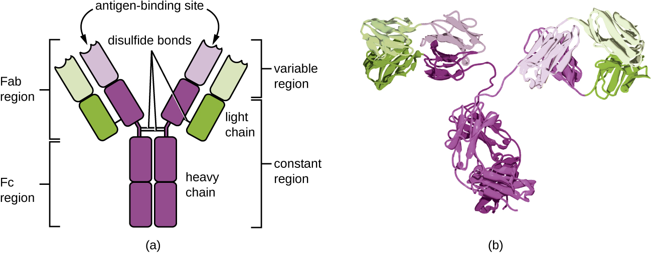
Two-panel diagram of an antibody. Left, a Y-shaped schematic with two purple heavy chains and two green light chains joined by disulfide bonds, antigen-binding sites at the tips of the two arms, and brackets marking the Fab and Fc regions and the variable and constant regions. Right, a ribbon model of the folded IgG protein in the same colors.
Figure 40.1: Structure of an IgG antibody. (a) Two heavy chains (purple) and two light chains (green) joined by disulfide bonds. The variable region at the tip of each Fab arm forms an antigen-binding site; the constant region makes up the rest of the arms and the Fc stem. (b) Three-dimensional structure. Figure 18.1.4 from Parker et al., Microbiology (OpenStax), Biology LibreTexts edition, CC BY 4.0 (Parker et al. 2016); panel (b) is a modification of work by Tim Vickers.

The two arms are the Fab regions, which bind antigen, and the stem is the Fc region; a flexible hinge joins them (Janeway et al. 2001b). The variable domains differ in amino-acid sequence from one antibody to the next, while antibodies of the same class and species have the same constant domains (Clark et al. 2018). At the tip of each arm, the variable domains of one heavy and one light chain together form the antigen-binding site (Janeway et al. 2001b). The variable tips therefore determine which target an antibody binds: binding depends on how well the site and the target match in shape and chemistry (Clark et al. 2018). The two arms are identical, so one IgG carries two copies of the same binding site.

The sandwich and the control line

The part of the antigen that an antibody binds is its epitope, and most protein antigens carry several different epitopes (Parker et al. 2016, sec. 20.1). A sandwich test holds the antigen between two antibodies (Koczula and Gallotta 2016). Both can attach to one antigen at once if they bind different epitopes. Two antibodies whose epitopes are the same or overlap block each other, and no sandwich forms.

The control line usually carries an antibody that binds antibodies from another species (Koczula and Gallotta 2016), such as an anti-mouse antibody when the detection antibody comes from a mouse. Because mouse antibodies of one class have the same constant domains, an anti-mouse antibody binds mouse antibodies of that class whatever their target. The control line therefore captures the detection antibody and its gold whenever the sample flows, with or without target.

ImportantKnowledge check

A developer proposes printing the control line with a second antibody against the viral protein. Would that line turn red for a sample with no viral protein, and what would the strip lose?

No. That antibody binds the target, not the detection antibody, so without target no gold collects there. The line would become a second test line, and a negative result could no longer be told apart from a strip through which the sample never flowed.

Where antibodies come from

Antibodies are made by B cells, white blood cells that each make antibodies with a single kind of binding site (Parker et al. 2016). A person carries at least \(10^{11}\) different antibodies (Janeway et al. 2001a). During an immune response, the B cells whose antibodies bind the foreign molecule most tightly are selected to multiply, so binding strength tends to rise as the response continues (Janeway et al. 2001a).

Monoclonal and polyclonal antibodies

Antibodies for a test are raised in an animal: a mouse, rabbit, or goat is injected with the target protein. Many of its B cells respond, each to its own epitope, and each multiplies into a clone: cells descended from one parent cell, all identical. The animal’s serum, the liquid part of blood after clotting (Betts et al. 2022), therefore holds a mixture of antibodies against different epitopes, called a polyclonal antibody (Parker et al. 2016, sec. 20.1). A monoclonal antibody comes from a single clone: every molecule has the same sequence and binds the same epitope (Table 40.1). In 1975 Köhler and Milstein fused B cells from an immunized mouse with tumor cells that keep dividing in culture. The fused cell, a hybridoma, keeps dividing in culture like its tumor parent and makes the one antibody of its B-cell parent (Köhler and Milstein 1975; Parker et al. 2016, sec. 20.1).

Table 40.1: Monoclonal and polyclonal antibodies (Parker et al. 2016, sec. 20.1).
Monoclonal Polyclonal
Source one B-cell clone, grown as a hybridoma many B-cell clones in an immunized animal
Epitopes recognized one several
Batch to batch uniform once the hybridoma exists composition varies

Monoclonal antibodies, mostly from mouse hybridomas, are preferred for lateral flow tests because one defined antibody can be produced in large quantities (Koczula and Gallotta 2016).

Ranking antibodies by affinity

An antibody’s binding site holds its epitope with a strength called its affinity (Clark et al. 2018). Affinity is quantified by the dissociation constant \(K_D\): the concentration of unbound antigen at which, at equilibrium, half of the antibody’s binding sites hold antigen. Its unit is moles per liter (M), and antibody values are given in nanomolar or picomolar, where \(1~\text{nM} = 10^{-9}\) M and \(1~\text{pM} = 10^{-12}\) M (Brown et al. 2020). The rate constant \(k_\text{on}\) measures how fast antibody and antigen bind and \(k_\text{off}\) how fast the complex dissociates (both defined in binding). Their ratio is \(K_D = k_\text{off}/k_\text{on}\).

Among antibodies that bind a suitable site without cross-reacting, a lower \(K_D\) means the antigen is held more tightly at equilibrium, since less antigen is needed to fill half the sites. The analytical sensitivity of a lateral flow assay is limited in part by the \(K_D\) of its antibody–antigen pair (Koczula and Gallotta 2016); the time to a visible line also depends on \(k_\text{on}\) (binding). A polyclonal antibody mixes binding strengths and has no single \(K_D\).

Example: antibody pairs and a polyclonal lot

A protein carries four epitopes, numbered 1 to 4, with a monoclonal antibody against each. Either antibody of a pair can serve as the capture antibody, so there are \(4 \times 3 = 12\) ordered (capture, detection) pairs. If epitopes 2 and 3 overlap, the pairs (2, 3) and (3, 2) fail, leaving 10. A test line printed with 2.0 µg of a polyclonal antibody of which 2.5% binds the target (illustrative values) carries \(0.025 \times 2.0~\mu\text{g} = 0.050~\mu\text{g} = 50\) ng of target-binding antibody.

Exercises

TipProblem 1

A viral protein carries six epitopes, numbered 1 to 6, with a monoclonal antibody available against each. Epitopes 1 and 2 overlap, as do 4 and 5: an antibody bound to one blocks the other.

  1. How many ordered (capture, detection) pairs of two different antibodies are there?
  2. How many of these fail because their epitopes overlap, and how many remain?
  3. A seasonal coronavirus protein also carries epitopes 3 and 6. Which remaining pairs would give a false test line for a person infected with the seasonal virus, and how many pairs are left without them?
  1. \(6 \times 5 = 30\).
  2. (1, 2), (2, 1), (4, 5), and (5, 4) fail, leaving \(30 - 4 = 26\).
  3. Only (3, 6) and (6, 3), for which both antibodies bind the seasonal protein. A pair such as (3, 1) captures the seasonal protein without labeling it. That leaves \(26 - 2 = 24\) pairs.
TipProblem 2

The test of Problem 1 is validated on 1,000 people without the target infection. Fifty of them carry the seasonal virus, and 4 of the other 950 give a false line for unrelated reasons. Design A uses the pair (3, 6), which gives a line for every seasonal carrier; design B uses (3, 1).

  1. Find the number of false positives and the clinical specificity of each design.
  2. Both designs have clinical sensitivity 0.85. At a prevalence of 5%, what is the positive predictive value (PPV) of each?
  1. Design A: \(FP = 50 + 4 = 54\), clinical specificity \(946/1000 = 0.946\). Design B: \(FP = 4\), clinical specificity \(996/1000 = 0.996\).
  2. With prevalence \(p = 0.05\), \(\text{PPV} = 0.85p/[0.85p + (1 - \text{spec})(1 - p)]\). Design A: \(0.0425/(0.0425 + 0.0513) = 0.45\). Design B: \(0.0425/(0.0425 + 0.0038) = 0.92\). With design A, more than half of the positive results are false, although the designs differ only in the epitope of one antibody.
TipProblem 3

A manufacturer compares polyclonal and monoclonal capture antibodies. The values below are illustrative, chosen for the calculation, not measurements.

  1. A test line is printed with \(1.0~\mu\text{g}\) of a polyclonal rabbit antibody. In lot 1, 3.0% of it binds the target; in lot 2, 1.2% does. How much target-binding antibody does each lot put on a strip?
  2. How much of lot 2 must be printed to match lot 1, and why does that still not make the lots equivalent?
  3. A hybridoma secretes a monoclonal capture antibody at 30 mg per liter of culture, and each strip uses \(0.40~\mu\text{g}\). How many strips does a 200 L culture supply?
  1. Lot 1: \(0.030 \times 1.0~\mu\text{g} = 30\) ng. Lot 2: \(0.012 \times 1.0~\mu\text{g} = 12\) ng, a factor of 2.5 less.
  2. \(30~\text{ng}/0.012 = 2.5~\mu\text{g}\). The lots also differ in which epitopes their antibodies bind and how tightly, so equal mass does not give equal binding.
  3. \(30~\text{mg/L} \times 200~\text{L} = 6.0~\text{g} = 6.0 \times 10^{6}~\mu\text{g}\), enough for \(6.0 \times 10^{6}/0.40 = 1.5 \times 10^{7}\) strips, all carrying the same antibody.
TipProblem 4

Candidate capture antibodies against the protein of Problem 1 have dissociation constants of 2.5 nM (A), \(4.0 \times 10^{-11}\) M (B), 300 pM (C), and \(8.5 \times 10^{-12}\) M (D). A fifth, E, is a polyclonal rabbit antibody.

  1. Express the \(K_D\) values of A through D in pM and rank the antibodies from tightest to weakest binding.
  2. By what factor do the tightest and weakest \(K_D\) values differ?
  3. Why can E not be ranked?
  4. The detection antibody binds epitope 6. A binds epitope 4, B binds epitope 2, C binds epitope 3, and D binds epitope 6. As in Problem 1(c), epitopes 3 and 6 also occur on the seasonal coronavirus protein. Which should be the capture antibody?
  1. A: 2500 pM, B: 40 pM, C: 300 pM, D: 8.5 pM. Tightest to weakest: D, B, C, A.
  2. \(2500/8.5 = 294\), about \(3 \times 10^{2}\).
  3. E mixes antibodies of different binding strengths, so it has no single \(K_D\).
  4. B. D binds the same epitope as the detection antibody, so the two block each other and no sandwich forms. C forms the pair (3, 6), which gives a false line for seasonal-virus carriers (Problem 1(c)). A and B both form valid pairs, since epitopes 4 and 2 do not overlap epitope 6 and do not occur on the seasonal protein, and B binds more tightly (\(K_D\) = 40 pM against 2500 pM).

References

Betts, J. Gordon, Kelly A. Young, James A. Wise, et al. 2022. Anatomy and Physiology 2e. OpenStax, Rice University. https://med.libretexts.org/Bookshelves/Anatomy_and_Physiology/Anatomy_and_Physiology_2e_(OpenStax).
Brown, Michael E., Daniel Bedinger, Asparouh Lilov, et al. 2020. “Assessing the Binding Properties of the Anti-PD-1 Antibody Landscape Using Label-Free Biosensors.” PLOS ONE 15 (3): e0229206. https://doi.org/10.1371/journal.pone.0229206.
Clark, Mary Ann, Matthew Douglas, and Jung Choi. 2018. “Antibodies.” 42.3 in Biology 2e. OpenStax, Rice University. https://openstax.org/books/biology-2e/pages/42-3-antibodies.
Janeway, Charles A., Paul Travers, Mark Walport, and Mark J. Shlomchik. 2001a. “The Generation of Diversity in Immunoglobulins.” In Immunobiology: The Immune System in Health and Disease, 5th ed. Garland Science, New York. https://www.ncbi.nlm.nih.gov/books/NBK27140/.
Janeway, Charles A., Paul Travers, Mark Walport, and Mark J. Shlomchik. 2001b. “The Structure of a Typical Antibody Molecule.” In Immunobiology: The Immune System in Health and Disease, 5th ed. Garland Science, New York. https://www.ncbi.nlm.nih.gov/books/NBK27144/.
Koczula, Katarzyna M., and Andrea Gallotta. 2016. “Lateral Flow Assays.” Essays in Biochemistry 60 (1): 111–20. https://doi.org/10.1042/EBC20150012.
Köhler, Georges, and César Milstein. 1975. “Continuous Cultures of Fused Cells Secreting Antibody of Predefined Specificity.” Nature 256 (5517): 495–97. https://doi.org/10.1038/256495a0.
Parker, Nina, Mark Schneegurt, Anh-Hue Thi Nguyen, Brian M. Forster, and Philip Lister. 2016. Microbiology. OpenStax, Rice University. https://bio.libretexts.org/Bookshelves/Microbiology/Microbiology_(OpenStax).